
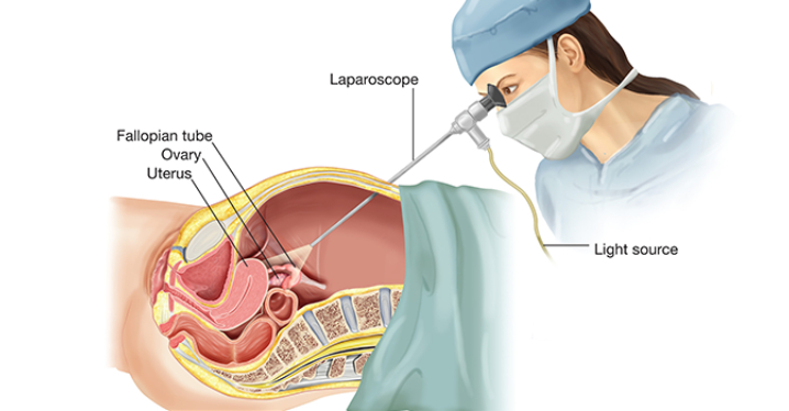
Diagnostic laparoscopy is a procedure that allows a doctor to look directly at the contents of the abdomen or pelvis.
How the Test is Performed?
The procedure is usually done in the hospital or outpatient surgical center under general anesthesia. The procedure is performed in the following way:
1)The surgeon makes a small cut (incision) below the belly button.
2)A needle or hollow tube called a trocar is inserted into the incision. Carbon dioxide gas is passed into the abdomen through the needle or tube. The gas helps expand the area, giving the surgeon more room to work, and helps the surgeon see the organs more clearly.
3)A tiny video camera (laparoscope) is then placed through the trocar and is used to see the inside of your pelvis and abdomen. More small cuts may be made if other instruments are needed to get a better view of certain organs.
4)If you are having gynecologic laparoscopy, dye may be injected into your cervix so the surgeon can view the fallopian tubes.
5)After the exam, the gas, laparoscope, and instruments are removed, and the cuts are closed. You will have bandages over those areas.
